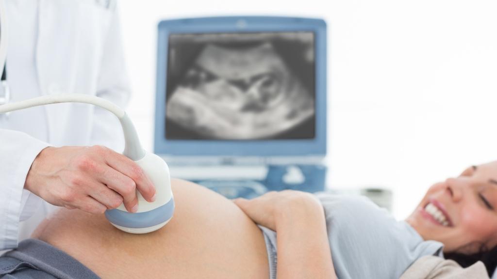
A study carried out by BCNatal and UPF opens the doors to the development of new therapies to improve lung development in underdeveloped fetuses

The study, carried out by the BCNatal Fetal Medicine Research Center (Hospital Clínic Barcelona and SJD) and the Unitat de Recerca BCN MedTech [BCN MedTech Research Unit] at UPF has discovered new scientific evidence that shows that problems with lung development in an underdeveloped fetus are related to vascular resistance. Using AI, computational models and ultrasound techniques, lung resistance of normal fetuses and underdeveloped fetuses have been analysed and compared when the mother receives additional oxygen therapy.
If, during pregnancy, fetal growth is lower than the normal threshold for each week of gestation, there is a higher risk that one of the baby's organs does not develop properly, which can negatively affect the newborn's health after birth.
The effect of restricted fetal growth on a person's health after birth and throughout their life has been the topic of several studies, especially with regard to brain and cardiovascular development. However, there is little evidence on how the lungs are affected.
This is the focus of this new study, jointly led by the BCNatal Fetal Medicine Research Center (Hospital Clínic Barcelona and SJD) and the Universitat Pompeu Fabra (UPF), which has allowed researchers to detect differences in lung development of fetuses with restricted growth versus normal growth, in terms of vascular resistance. The study was carried out by measuring fetal blood flow velocity and analysing it using artificial intelligence and computational models.
The results of the study, recently published in an article in the Scientific Reports (Nature) journal, open the doors to the development of new therapies to improve lung development in underdeveloped fetuses and to preventing respiratory health problems that could persist well into infancy, and even into adolescence and adulthood.
The principal investigators of the study are Fàtima Crispi—investigator at BCNatal and part of the Fetal and Perinatal Medicine team at Clínic-IDIBAPS—and Bart Bijnens (ICREA, UPF), investigator at the BCN MedTech Research Unit in the Department of Engineering at UPF. The other investigators come from various departments and research groups at Clínic-IDIBAPS, and are also affiliated with the University of Barcelona and CIBER networks for respiratory diseases and rare diseases.
More than 200 pregnant women took part in the study
This investigation looked at fetal blood flow velocity and how it was affected when the mother received supplementary oxygen, with 208 women taking part, all between 24 and 37 weeks pregnant. Each participant attended Hospital Clínic Barcelona, where they underwent all the relevant testing for this study. In 97 cases, the fetus showed restricted growth, which resulted in a very low birth weight, and in 111 cases, the fetus showed normal growth. For each fetus, the blood flow velocity was measured in the main arteries and pulmonary vessels, and results were compared using AI. Plus, lung resistance was calculated using a computational model.
The pulmonary blood flow velocity of the fetuses was analysed in two conditions: one when the mother was breathing normally; the other when the mother was given supplementary oxygen through a face mask (i.e. in a hyperoxygenated state). This analysis was done using a fetal ultrasound technique to estimate blood flow velocity.
That said, organ resistance, such as in the case of the lungs, cannot be directly measured with ultrasound, therefore requiring a computation model to represent the heart and blood vessels. To better understand this concept, we could compare this computational model with the simulation of an electronic circuit. Researchers digitally recreated the fetal blood vessels and, using real blood flow velocity measurements and simulating the rest, they were able to estimate both the resistance and elasticity of various organs.
Blood flow velocity and vascular resistance in the lungs of the fetuses with restricted growth were able to be normalised by providing supplementary oxygen to the mother
Finally, the investigation team used automated learning models based on artificial intelligence techniques to compare fetal blood flow patterns, thereby allowing them to categorise them according to flow pattern and clinical parameters. In subsequent analyses of the effects of hyperoxygenation, researchers were able to link changes in fetal lung resistance to oxygen supplementation of mothers, demonstrating that higher oxygen concentrations improved blood flow to the lungs of fetuses with restricted growth, while having no effect on normally growing fetuses. ‘Essentially, the results of our study show that, in restricted-growth fetuses, mean blood flow velocity and vascular resistance in the lungs are different to those of normal-growth fetuses, and that this difference can be normalised by providing the mothers with additional oxygen,’ explains Bart Bijnens (ICREA, UPF).
‘Detecting these differences in the vessels of the lungs opens the doors to developing new therapeutic strategies to improve lung function in restricted-growth fetuses. These improvements during fetal development may reduce the risk of respiratory diseases after the child is born,’ adds Dr Fàtima Crispi (BCNatal, Hospital Clínic).


