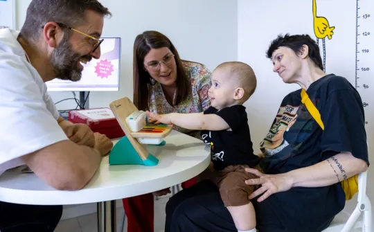
In BCNatal - SJD Barcelona Children's Hospital we are specialists in the treatment of spina bifida and in the precise fetal surgery for its treatment.
What is spina bifida?
Spina bifida (myelomeningocele) is a birth defect in which the skin and bone of an area of the spine have not formed properly and a part of the spinal cord, which is normally protected within the spine, is exposed. Therefore, throughout pregnancy the spinal cord is exposed to amniotic fluid and deteriorates, worsening its function before birth.
The spinal cord contains the nerves that carry information from our brain to the rest of the body, so at birth symptoms such as decreased movement of the legs, loss of sensation and problems with the control of urine or faeces appear. This complication occurs in 1 in 2000 pregnancies.
Why does it occur?
The main cause of spina bifida is a lack of folic acid, either due to an inadequate diet of the pregnant woman or due to alterations in her metabolism.
In some people with genetic susceptibility, this lack of folic acid causes a failure in the closure of the spine. In fact, the risk of recurrence after having a baby with spina bifida is 4-5 %, so in that case it is advisable to take folic acid at high doses already 3-4 months before pregnancy.
Some medications for epilepsy, as well as obesity or diabetes of the pregnant woman, are also considered risk factors for neural tube defects.
Although there is genetic susceptibility, it is not possible to find an abnormal gene or chromosome, so most children with spina bifida do not have genetic alterations. In a small proportion, spina bifida can be accompanied by genetic problems, so a study is advisable and is usually carried out by amniocentesis.
What problems will a child with spina bifida have?
The exposure of the spinal cord to amniotic fluid causes it to be damaged throughout pregnancy, with neurological problems, both motor and sensory, appearing below the level of the injury. In the case of defects at the level of the chest or upper lumbar area there is very often paralysis of the legs and it is not possible to walk; a wheelchair is often needed.
However, most defects occur in the lower back, the lumbo-sacral area, and in these cases walking is usually possible, usually with help (crutches, cane). The sensitivity below the lesion is reduced, and sphincter problems (incontinence and retention of urine and/or faeces) as well as those of sexual function also appear in a large proportion of cases. There may also be problems of malposition of the feet, such as talipes, or of knees and hips. In short, the higher in the spine the defect, the more severe the complications.
In addition, open spina bifida is associated with Arnold-Chiari malformation type II, which consists of a displacement of the cerebellum into the cervical spine so that the normal flow of cerebrospinal fluid is obstructed. This causes an increase in fluid in the brain (hydrocephalus) which in turn can cause an increase in intracranial pressure and significant secondary damage.
Therefore, it is important to drain the fluid after birth by placing a small tube or shunt, which improves brain pressure but can have complications and overall worse cognitive outcomes.
Diagnosis
The severity of motor and sensory symptoms will depend mainly on the level of spinal cord injury and the presence of fluid within the brain (hydrocephalus).
Once the diagnosis has been made, a comprehensive evaluation by an experienced fetal medicine team is essential, which will assess whether it is an isolated spina bifida, that is, whether there are any other malformations or associated genetic abnormalities, and the level of the spinal cord injury, that is, which vertebra is affected.
To this end, a series of studies and different assessments are carried out:
- Specialized ultrasound, to study in detail the brain and spine to assess the level of the lesion and the presence of hydrocephalus.
- Magnetic resonance imaging.
- Amniocentesis and genetic study.
- Multidisciplinary assessment involving specialists in fetal medicine, neonatologists and paediatric neurosurgeons.
Treatment
Once the study is complete, all the details are discussed at length with the parents, who always have the final say on treatment.
Conventional treatment consists of repair and closure of the lesion within the first 24–48 hours of birth. In addition, it is usually necessary to place a shunt for severe hydrocephalus (for more information see 'What problems will a child with spina bifida have?').
Since spinal cord damage is related to its exposure to amniotic fluid, the possibility of treating it during fetal life, to prevent the prognosis from worsening after birth, has become a treatment option in some cases. In fact, there is evidence that performing fetal surgery in some cases (depending on the level of the lesion and the degree of hydrocephalus) reduces the need to place a shunt at birth and improves motor function.
The fetal surgery that has shown benefit in the evolution of the disease is that performed between the 19th and 26th weeks of pregnancy. It consists of accessing the fetus and repairing the defect in the back.
In SJD Barcelona Children's Hospital, an incision is made in the pregnant woman like that of a caesarean section. Another incision is made in the womb through which we access the fetal back and an expert neurosurgeon closes the defect with layers to protect the spinal cord. In our centre we make a small (less than 5 cm) incision in the uterus which greatly reduces the risk of complications. It is a surgery that requires continuous monitoring of the pregnant woman and is associated with a risk of prematurity.
Some centres in the world also offer an endoscopic repair, that is, the same incision is also made in the skin of the pregnant woman, but the entry into the uterus is done with endoscopy. After a two-year work in experimental models, in BCNatal we have decided to continue with the incision in the uterus, since it allows a much more precise repair of the defect in the back, and a much shorter surgery time at the moment.
In any case, you can always evaluate the type of surgery, depending on the characteristics of the case, and what is considered to represent a faster and less aggressive surgery for the pregnant woman and her uterus, and of course for the fetus.
What controls are needed for fetal surgery?
Before surgery, parents can discuss in detail with paediatric specialists all the details of postnatal treatment, what they can expect and what the quality of life of babies with this problem is like.
The in-patient admission of the pregnant woman will initially last 4-5 days; afterwards weekly check-ups with examination and ultrasound will be required, and she will have to maintain a low-activity lifestyle mainly at home until the end of pregnancy, especially for the first 3-4 weeks after the intervention. Normally, at around 37 weeks gestation is ended with a caesarean section.
During pregnancy, you will receive the support of nurses specialized in fetal medicine, not only on a technical level but also on an emotional level throughout the process. In addition, we can put you in contact with other families who have gone through the same experience. This is very positive and helps to humanize and understand the problem in a much more intuitive way and without the difficulties that sometimes arise when receiving only technical information from professionals.
Why BCNatal - SJD?
For parents who wish to continue their pregnancy care and have their baby with us, we offer a fetal surgery team with the best survival and quality of life figures that can currently be obtained.
Our fetal surgery team offers the best survival and quality of life figures that can currently be obtained.
Experience and efficiency
Our fetal surgery team offers the best survival and quality of life figures that can currently be obtained.
- We are among the centres with the most extensive experiences in fetal surgery in the world, with more than 2000 interventions performed, the shortest surgical times reported in the medical literature and, consequently, very low rates of complications.
- In addition to our substantial surgical experience, being at the same time one of the most important research and development centres at an international level, we constantly incorporate improvements in materials and techniques that allow us to further increase the precision and speed of our interventions.
We are a multidisciplinary team, which allows us to approach surgery in a comprehensive way: from the choice of the best strategy to individualized postnatal follow-up.
Highly specialized team
We are a multidisciplinary team, which allows us to approach surgery in a comprehensive way: from the choice of the best strategy to individualized postnatal follow-up.
In the specific case of spina bifida, fetal surgery is carefully prepared and simulated with 3D models by a multidisciplinary team made up of fetal surgeons and neurosurgeons. The Neurosurgery Department at SJD Barcelona Children's Hospital has extensive experience in the repair of spina bifida and is capable of performing a high-precision repair with the best possible results.
Specifically, babies with spina bifida will be known and evaluated by our high-level Neonatology, Neurology, Neuroradiology and of course Neurosurgery departments before they are born. The Hospital has highly qualified medical and nursing professionals caring for these delicate patients and attending to their needs 24 hours a day, 365 days a year.
We accompany you with an individualized postnatal follow-up.
After the birth we continue with our patients
We accompany you with an individualized postnatal follow-up.
To the excellence of the prenatal team is added a third-level paediatric centre with teams made up of a large number of paediatric cardiologists who have the best and most modern technology.
Once discharged, our Paediatric teams will follow the baby for the first few months, and then years and take care of him or her to achieve optimal development and to solve any problems in this very fundamental part of life.
